Pilbarascutigera incola (Verhoeff, 1925)
Description
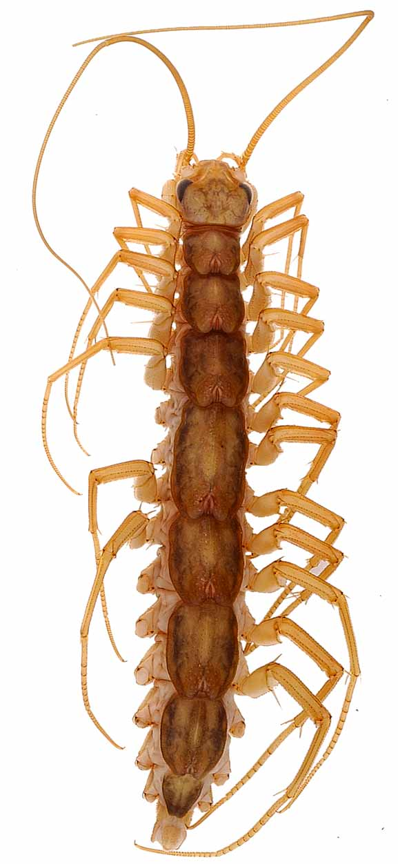
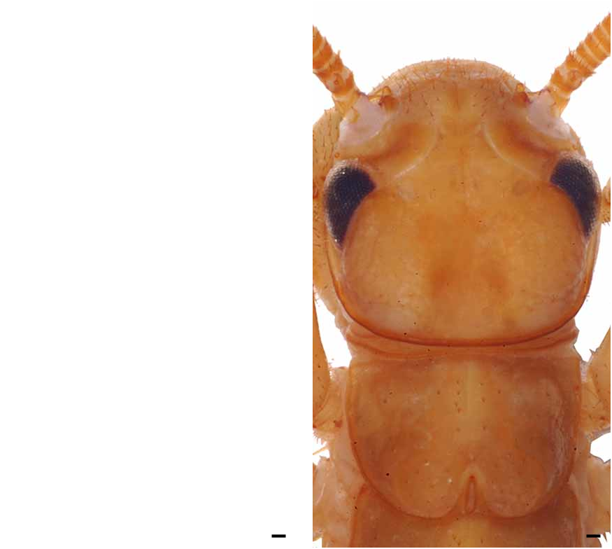
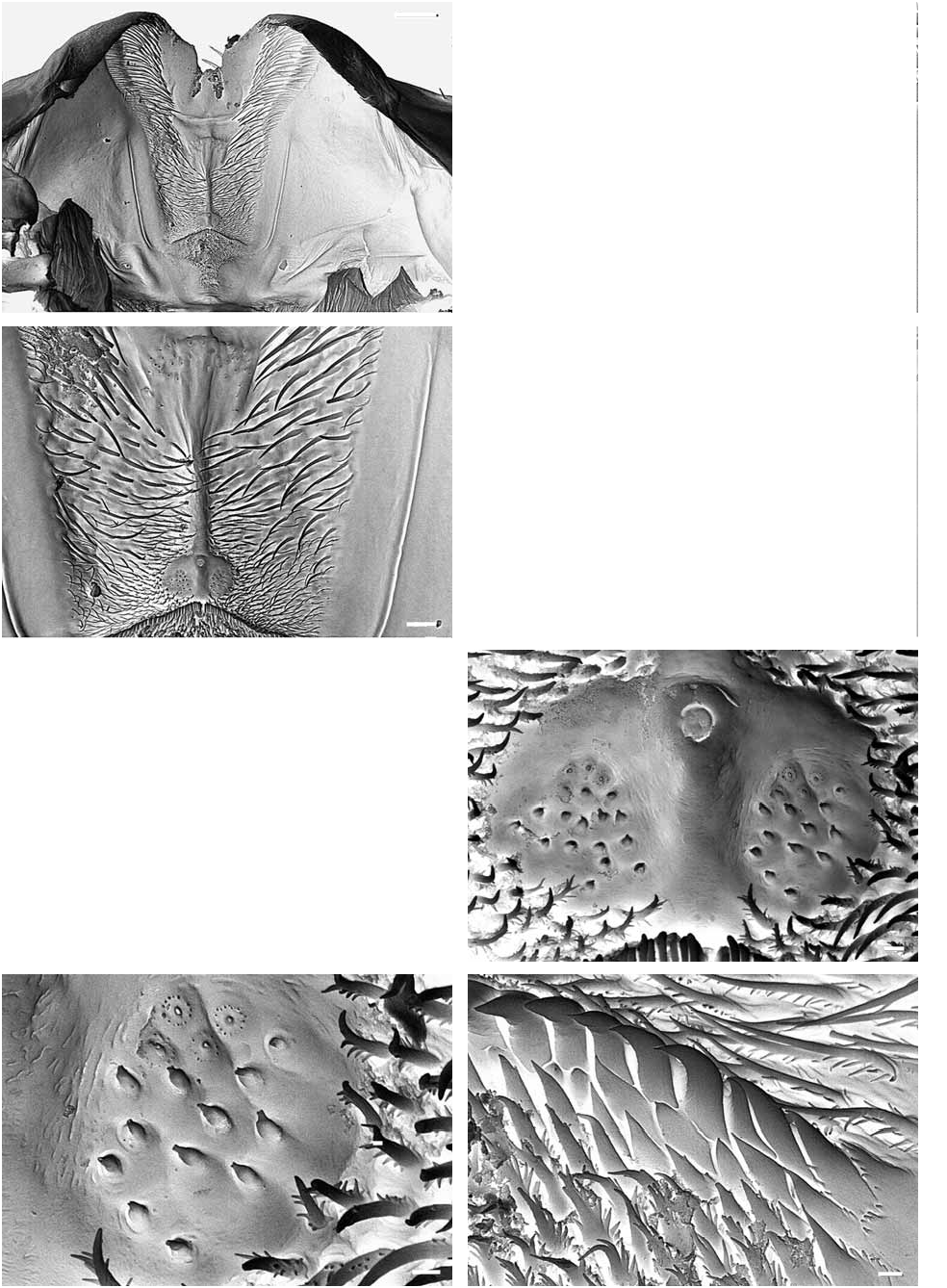
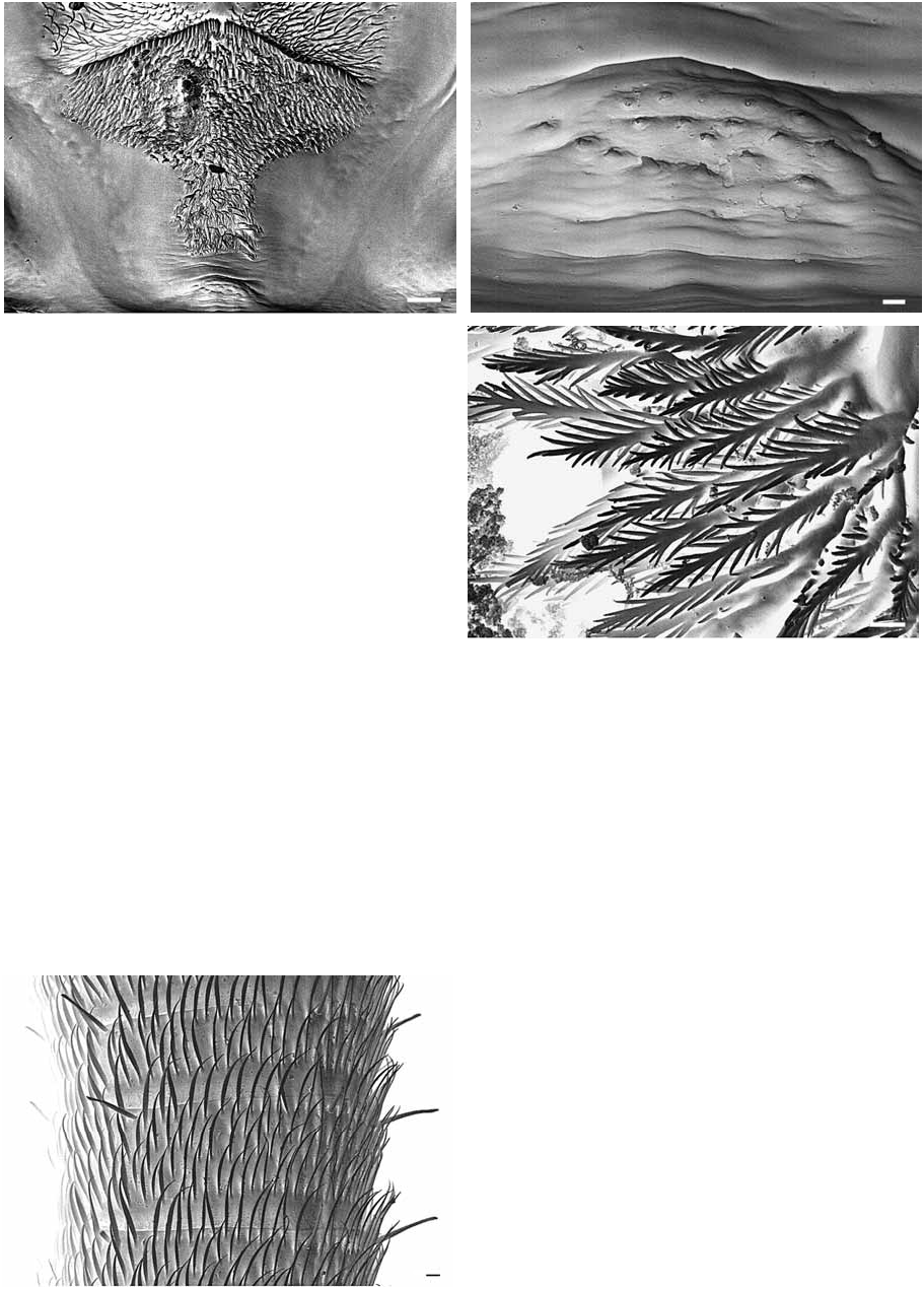
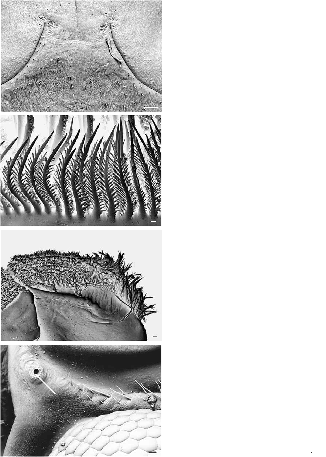
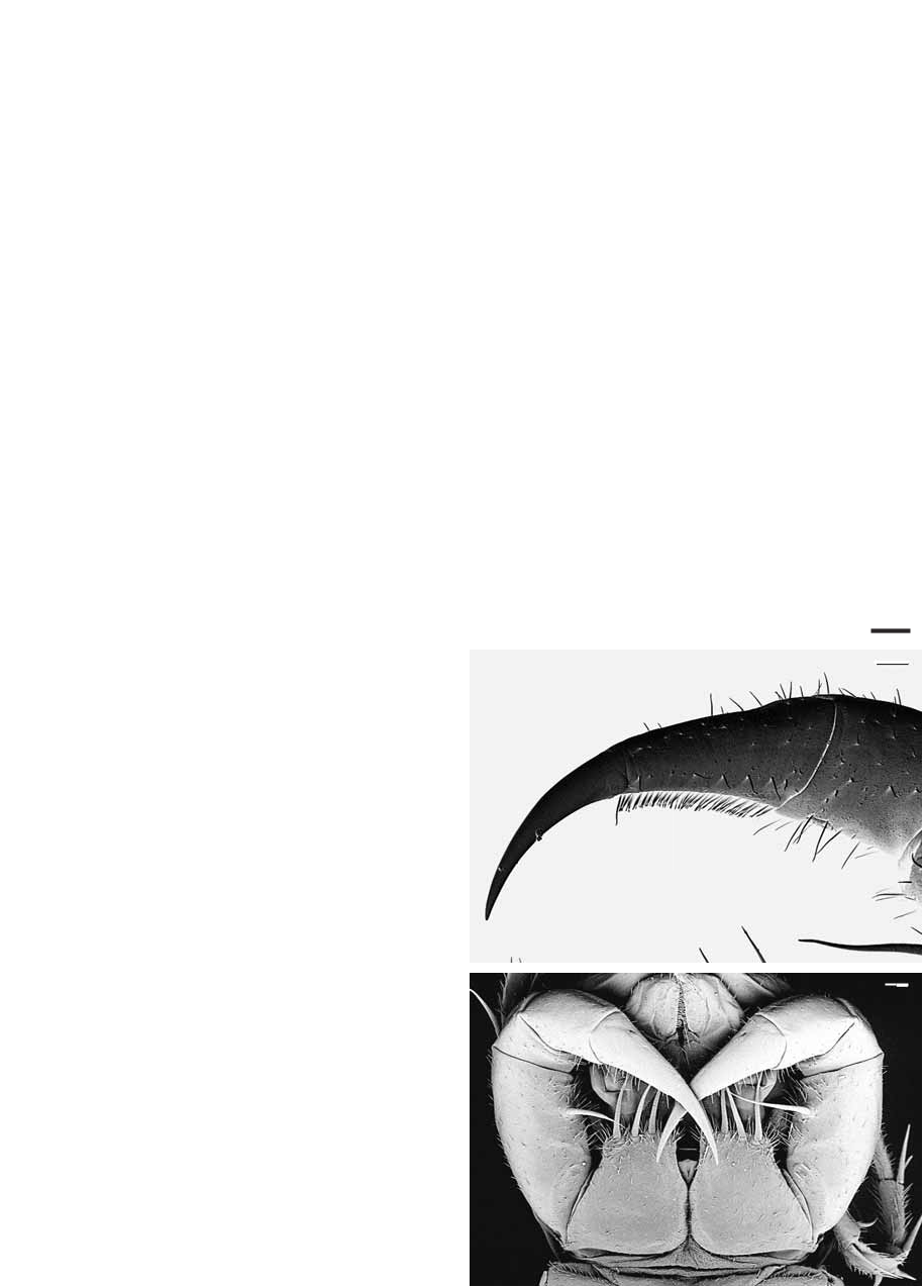
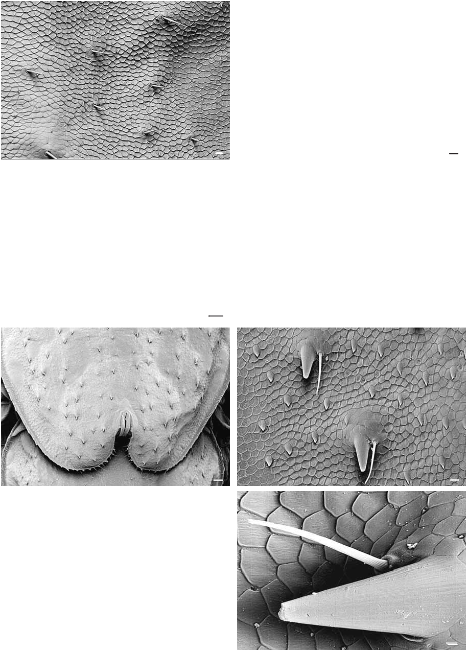
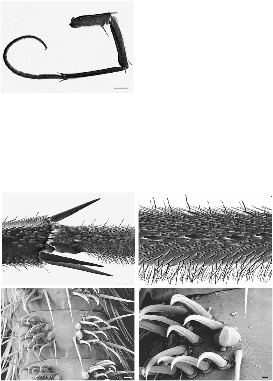
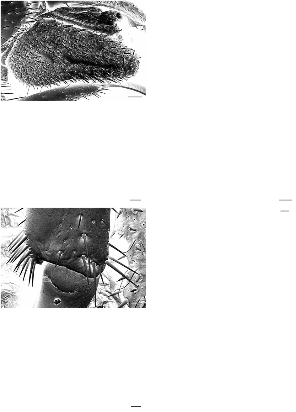
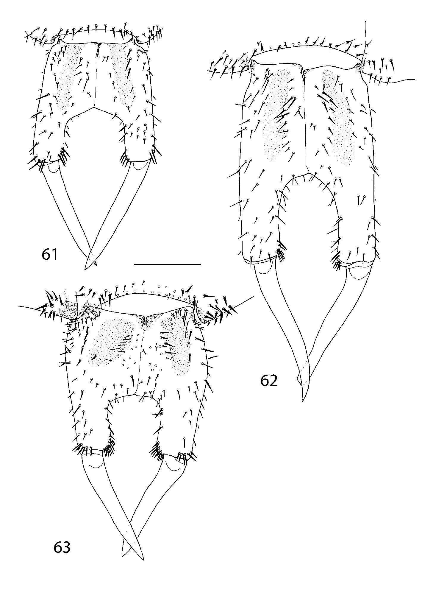
Description: Length up to 31 mm in males, 28 mm in females. Colour: Head capsule typically orange-brown with radiating dark brown network covering most or all of dorsal side of head behind sutures; some populations with rather uniformly orange head capsule, radiating network of darker pigment confined to posterolateral part of head; tergal plates with yellowish-orange, bright orange or pale tan medial band with diffuse dark brown, blackish or orange-brown pigment that becomes more concentrated as a pair of longitudinal bands on more posterior segments; stoma saddles generally orange-yellow or pale tan / whitish, often with diffuse blackish, red-brown or reddish pigment; lateral flanks of tergal plates pale orange, each side with a variably strong mottled longitudinal band of blackish, dark brown or reddish-brown pigment. Legs pale to moderate orange, sometimes yellow, usually without significant concentric banding, rarely with well defined bluish bands on femur, tibia, and basal article of tarsus I. Sternites yellow-orange in anterior segments, more intensely orange in posterior segments. Head capsule: Anterior projection of sutures short, triangular, pointing straight anteriorly (Fig. 21). Posteromedian impression moderately deep. Antenna 1.2 – 1.5 times body length. First flagellum with 56 – 124 articles apart from specimens in which node is undifferentiated with extreme asymmetry (e. g., 65 / 173 or 174 / 78 articles) between left and right antennae, mean 75 articles (N = 84, specimens> 14 mm body length); asymmetry between numbers of articles in first flagellum on each side of a specimen usually less than 10 articles, ranging to 26; ring-like articles with two or three whorls of densely arranged hairs with a flattened section and gentle curvature (Fig. 19) and a few trichoid setae in a single whorl encircling distal end of article (Fig. 20); hairs on distal part of second flagellum in either one or two whorls per article, these differing whorl numbers typically on alternating articles. Tömösváry organ small, in typical position between eye and base of antenna (Fig. 27); median pore encircled by a few rings of small swellings. Epipharynx: Lateral bar of labral trapezoid with narrow longitudinal groove along whole length of bar (Fig. 7). Labral bristles differentiated into narrow outer band of short, pectinate bristles and wider inner band of longer simple bristles (Fig. 6). Two clusters of sensilla along midline of labral trapezoid: more distal unpaired, transverse group of a few dozen bottle-shaped sensilla (Fig. 9) at termination of median ridge (Fig. 7), and proximal cluster composed of three aggregations of sensilla at proximal sclerotized bulge (Fig. 10); proximal aggregations comprise a lateral pair of groups of mostly bottle-shaped sensilla and a few buttonshaped sensilla (Fig. 11), and slightly distal to the lateral aggregations a median side-by-side pair of buttonshaped sensilla. Chevron-shaped spine row of triangular and distally-curved denticles at border between labral and clypeal part of epipharynx (Fig. 12); immediately proximal to spine row is dense median field of pectinate spines composed of broadly rhomboid distal portion with pectinate spines and narrow, subparallel-sided proximal portion (Fig. 13) with multifurcating spines. Broadly ovate cluster of nipple-and bottle-shaped sensilla on medial part of clypeal triangle (Fig. 14), a short distance behind dense field of branching spines (Fig. 13); ovate groupings of about 10 nipple-shaped sensilla form pair of lateral clusters set in weak depression at a level of about midlength of dense median spine field. Pair of lateral spine fields (Fig. 15) proximal to lateral clusters of sensilla within clypeal part of epipharynx; spines uniformly slender, elongate; a few pores of apparently epidermal glands in spine field. Median transverse band of six or seven spines, some bifid, immediately in front of mouth. Hypopharynx: Elongate, deeply projecting into preoral chamber (Fig. 17). Bars of lateral sclerotised fork with lateral bulges. Median excavation on proximal part of frontal surface bordered laterally by flattened bipectinate bristles (Fig. 16); pectinate bristles form in addition a few rows on distal part of hypopharynx (Fig. 18). Paramedial rows of button-like sensilla on tip of tongue, each sensillum with an ovate rim that is in turn enclosed by an incomplete, horseshoe-shaped rim (Fig. 18, inset). Cluster of sensilla-like structures in front of mouth between median excavation and converging flattened bars of proximal fork with more or less rhomboid arrangement. Mandible: About 20 pectinate lamellae in large specimens (Fig. 22); individual spines at tips of pectinate lamellae with ridge on inner side and many small, blunt marginal serrations. Three teeth, each with three cusps, surface smooth (Fig. 24). Narrow fringe of pectinate bristles along dorsal half of pectinate lamellae and against teeth; each bristle with many short lateral branches along all but terminal part (Fig. 23). Haarpolster has narrow molar plate along most of its length (Figs. 25, 26); surface of molar plate composed of about six dense spine rows. Haarpolster bristles arranged in numerous imbricated bands of pectinate bristles. First maxilla: Dense field of brush-like setae on inner margin of distal article of telopodite with typical scutigeromorph structure (basal half smooth, distal half with many short back-curved spines: Fig. 28). Coxal process with short, simple setae evenly scattered over its ventral surface. Second maxilla: Typical scutigerid arrangement of elongate spine bristles at distal ends of podomeres (dorsal and ventral spine bristles on prefemur; four spine bristles on femur; two dorsal spine bristles on tibia: Figs. 29 – 31). All spine bristles with similar surface details, spiniform scales, short proximally, becoming more elongate distally (Fig. 32) to confer a ridged or fluted surface to distal third of spine bristle. Tarsus densely setose, with slender, distally curved, setiform spines between longer trichoid setae at tip (Fig. 33). Maxillipede: Four spine bristles on anterior margin of coxa about equal in length to coxa (Fig. 36); surface ornament of spine bristles similar to those on second maxilla. Spine comb (Dornenkamm of Borucki 1996) along inner side of tarsus (Fig. 34); spines twisted distally, terminating in a rounded bulb (Fig. 35). Poison calyx cylindrical; poison duct extending to base of tarsus. Tergal plates: Outlines as shown in Fig. 2. Stoma saddles weakly inflated. Spiracles relatively short (Fig. 41). Spines first appear on T 3 in most specimens, exceptionally T 2, in which case they are few and confined to the lateral part of the tergal plate or include one to three spines on stoma saddle; spines consistently paired with a Stachelborste or, more commonly, a Tastborste; pairing of spine and Stachelborste most common on anteromedian part of TT 3 – 4, with more posterior parts of those plates having spines paired with Tastborsten; consistent pairing of Tastborste and spine on TT 5 – 8 (Fig. 44); Tastborsten generally slightly longer than their associated spine. Numerous isolated Tastborsten (not associated with spines) on tergal plates (Fig. 40). TT 4 – 7 with more or less even distribution of spines / Tastborsten on medial and lateral parts of tergal plate as well as on stoma saddles (Fig. 41). Spines usually first appear on stoma saddle of T 3 or T 4, sometimes on T 5, rarely on T 2 stoma saddle; number of spines on stoma saddles as shown in Table 1. Spicula small, triangular, each separated by several polygonal scales that lack spicula (Fig. 43), in some specimens spicula weaker on medial than lateral parts of tergal plates. Margins of TT 1 and 2 with alternating Tastborsten and Stachelborsten; spines fringe margin from posterior ½ – 2 / 3 of T 3 – or T 4 – T 8; marginal spines quite varied in size, from small to moderately large, each spine paired with a Tastborste (Fig. 43). Legs: Tarsus I and II segmentation as shown in Tables 2 and 3. Leg 15 with up to 359 tarsomeres. Prefemoral spine bristles (“ end spines ”) 2 / 1 on legs 1 – 14, 1 / 1 on leg 15. Femoral spine bristles 1 / 2 on all legs. Tibial spine bristles 0 / 1 on leg 1, 1 / 1 or ½ on leg 2 (posterior ventral spine bristle short when present), consistently ½ from leg 3, posterior ventral spine bristle becoming about as long as anterior ventral spine bristle (Fig. 49) from leg 5. Spine rows on prefemur (Fig. 47), femur (Fig. 48) and tibia consistently with spine / Tastborste pairing. Basal article of tarsus 1 with spines (single distal posterior spine) first appearing on legs 3 – 6, irregularly increasing on more posterior legs to maximum of 8 – 16 spines in longitudinal posterior row in leg 14 (Fig. 50); numbers on each leg variable (e. g., leg 8 with 3 – 7 spines in longitudinal posterior row). Tarsus II of legs 1 – 14 with tarsal papillae (Tarsalzapfen) on successive segments excepting first and last few segments on each leg; papillae usually paired, exceptionally arranged as two pairs per segment (Fig. 51). Cluster of setae on ventrolateral side of tarsi II arranged in two or three transverse bands of three or four setae per band (Fig. 52). Resilient sole hairs (federnden Sohlenhaare of Verhoeff 1904 b) originating near posteromedial edge of tarsal papilla, extending to about half length of the succeeding segment. Sternites: Posterior margin with gentle to moderate concavity medially, on each side of a posteriorly-convex extent. Longitudinal median furrow in anterior half or more of sternites; sternites scattered with setae except for on posterior part of anterior segments; margin fringed by many short setae; sternal setae finer on more posterior segments, which bear minute multifurcating spinulae (Haardörchen of Verhoeff) and scattered Tastborsten. Female: Gonopod with maximum length 1.7 – 2.5 times maximum width (ratio A / B of Würmli 1973, fig. 1); longitudinal median suture in proarthron complete. Lateral margins of proarthron gently divergent posteriorly (Fig. 61), nearly parallel sided (Fig. 62), or gently convergent posteriorly (Fig. 63). Subtriangular depression (Grube of Verhoeff) on proarthron shallow, bearing at most just a few short setae (Fig. 56). Proarthron 1.0 – 1.45 times length of mesarthron (ratio C / D of Würmli 1973). Coarsest setae on proarthron slightly thicker than those on mesarthron apart from cluster at distomedial corner of mesarthron, this cluster usually composed of 10 – 15 setae (Fig. 57). Sinus between mesarthron usually broadly parabolic, its apex typically weakly pointed but sometimes distinctly pointed (Fig. 61) or, less commonly, approximately transverse (Fig. 63); width of mesarthron 0.5 – 0.8 maximum width of sinus (ratio F / G of Würmli 1973). Mesarthron with numerous sensilla (apparently sensilla coeloconica; cf. Ernst & Rosenberg 2003) between its setae, including conical sensilla (Fig. 58, inset); sensilla surrounded by narrow, circular rim. Proarthron + mesarthron 1.0 – 1.7 times length of metarthron (ratio C + D / E of Würmli 1973). Outer margin of metarthron nearly straight for much of its length. Ventral surface of metarthron scattered with small, blunt sensilla coeloconica (Fig. 59); single row of sensilla coeloconica along outer margin of metarthron; a few setae on dorsal side of metarthron. Subanal plate drop-shaped, maximum height slightly more than half its length, with blunt, rounded distal end and nearly symmetrical dorsal and ventral margins (Fig. 53); smooth, non-setose band along middle of subanal plate in its posterior half to two-thirds (Fig. 54); setae of varied size on outer surface of subanal plate, predominantly slender subspiniform setae and short, conical, spiniform setae, between which are slender, curved hairs (Fig. 55); setation becoming coarser posteroventrally, with spiniform setae concentrated on posterior third of subanal plate. Telson elongate triangular with rounded posterior apex in both sexes, bearing abundant setae and slender, curved hairs as on subanal plate. Male: Typically scutigerid gonopod styles on first and second genital segment (Fig. 60); both pairs of gonopods densely covered with setae; terminal part with short, spiniform setae between longer setae. Subanal plate with parabolic outline, relatively shorter than in female, non-setose band along its length at midwidth.
Common Names
No common names available.
Taxonomic Hierarchy
Kingdom: Animalia
Phylum: Arthropoda
Class: Chilopoda
Order: Scutigeromorpha
Family: Scutigeridae
Genus: Allothereua
Species: Pilbarascutigera incola (Verhoeff, 1925)
Pilbarascutigera incola (Verhoeff, 1925)